17-Hydroxyprogesterone ELISA Kit
Number of Plates
- Catalog Number K053-H
- Assay Type Competitive ELISA
- Sample Types Extracted Serum, Extracted Plasma, Urine, Fecal Extracts, Tissue Culture Media
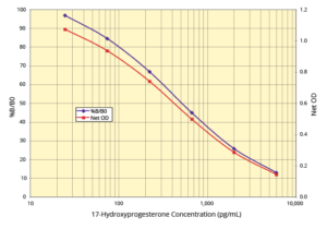
- Sensitivity 20.3 pg/mL
- Species 17HO-P is identical across species
- Assay Duration 1.5 Hours
- Samples/Plate 40 in Duplicate
- Readout Colorimetric, 450 nm
Personal Touch
Here to Help.
Ready to Ship
Most kits in stock.
Easy to Use
Simple protocols.
Assay Principle:
The 17-Hydroxyprogesterone ELISA Kit quantitatively measures 17-Hydroxyprogesterone (17HO-P) in extracted serum, extracted plasma, urine, fecal extracts, and tissue culture media. This competitive ELISA has a run time of 1.5 hours. Please read the complete kit insert for more information before performing this assay. Use our provided 17HO-P standard to generate a standard curve for the assay.
Protocol Summary:
- Introduce standards or diluted samples into the provided transparent microtiter plate, coated with donkey anti-sheep IgG antibody.
- Add 17HO-P peroxidase conjugate and 17HO-P polyclonal sheep antibody to initiate the immunological reaction.
- Incubate the mixture covered at room temperature, shaking for 1 hour. The reaction inversely correlates with the 17HO-P concentration in the sample.
- After incubation, remove excess conjugate and introduce the TMB substrate. This substrate reacts with the bound conjugate to generate a colorimetric signal.
- Measure the signal intensity using a plate reader at 450nm and calculate the 17HO-P concentration based on the standard curve.
Background:
17-Hydroxyprogesterone (17HO-P) is a steroid hormone in the androgen group found across various species including mammals, reptiles, and birds. It was first isolated by Pfiffner and North in 1940. Produced primarily in the adrenal glands and the corpus luteum of the ovary, 17HO-P serves as a precursor in the biosynthesis of cortisol. It undergoes hydroxylation at the 11 and 21 positions to form cortisol.
Deficiencies in enzymes like 11- or 21-hydroxylase lead to reduced cortisol synthesis, disrupting the feedback inhibition of adrenocorticotropic hormone (ACTH). This results in elevated production of 17HO-P. Conversely, deficiencies in 17-alpha-hydroxylase or 3β-hydroxysteroid dehydrogenase type 2 can lead to lower 17HO-P levels, with potential increases in progesterone or pregnenolone, respectively.
Normal levels of 17HO-P vary based on age and gender, with specific ranges established for children (3-90 ng/dL) and women in different phases of the menstrual cycle (20-100 ng/dL before ovulation and 100-500 ng/dL during the luteal phase). The DetectX® 17-Hydroxyprogesterone ELISA Kit is an important tool for endocrinologists and researchers, providing a sensitive and specific method for measuring 17HO-P levels in a range of biological samples. This kit facilitates the study of steroid hormone biosynthesis, adrenal function, and related physiological processes.