Glucose Fluorescent Detection Kit
The DetectX® Glucose Colorimetric Detection Kit is designed to quantitatively measure glucose in a variety of samples.
Personal Touch
Here to Help.
Ready to Ship
Most kits in stock.
Easy to Use
Simple protocols.
- Catalog Number K039-F1
- Assay Type Detection Kit
- Sample Types Serum, Plasma, Urine, Buffers, Tissue Culture Media
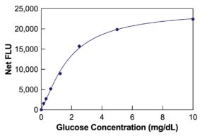
- Sensitivity 8.24 µg/dL
- Species Species Independent
- Assay Duration 30 Minutes
- Samples/Plate 40 in duplicate
- Readout Fluorescent, 590 nm emission / 520 nm excitation
Assay Principle:
The DetectX® Glucose Fluorescent Detection Kit quantitatively measures Glucose levels in serum, plasma, urine, buffers, and tissue culture media. This detection kit features a 30-minute run time for quick and accurate analysis. Read the complete kit insert for detailed instructions. The kit includes a Glucose standard essential for establishing an accurate standard curve.
Protocol Summary:
- Place standards or diluted samples into the provided black microtiter plate.
- Add Substrate, Horseradish Peroxidase Concentrate, and Glucose Oxidase Concentrate to each well, ensuring even mixing of reagents.
- Incubate the mixture at room temperature for 30 minutes. During this time, glucose oxidase reacts with glucose to produce hydrogen peroxide, which then reacts with the substrate in the presence of HRP to generate a fluorescent product.
- After incubation, use a plate reader to detect the fluorescent signal at 590nm. Determine the glucose concentration using the intensity data and the standard curve.
Background:
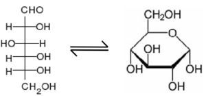
Glucose is a fundamental carbohydrate and primary energy source for cells. As a monosaccharide, aldose, hexose, and reducing sugar, it has a central role in energy metabolism across a wide range of biological systems. Glucose regulation and metabolism are critical to maintaining cellular homeostasis and integrity. Fluctuations in glucose levels can have significant biological implications.
Hypoglycemia, or a severe drop in blood glucose, can lead to metabolic dysfunction, neuroglycopenia, seizures, and even death. Conversely, chronic hyperglycemia, or elevated blood glucose levels, contribute to “glucose toxicity,” which can lead to β-cell dysfunction and various complications commonly associated with diabetes. Moreover, estrogen-induced signaling pathways in neurons involve mitochondria to enhance mitochondrial function, supporting key processes like aerobic glycolysis, citric acid cycle, oxidative phosphorylation, and ATP generation.
The DetectX® Glucose Fluorescent Detection Kit is an essential tool in research and clinical laboratories. It enables the precise quantification of glucose in various sample types, supporting studies in cellular energy metabolism, diabetes research, neurobiology, and other fields where glucose measurement is crucial.