Assay Principle:
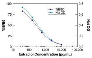
The DetectX® Estradiol ELISA Kit quantitatively measures estradiol in urine, dried fecal extracts, and tissue culture media. This competitive ELISA, with a run time of 2.5 hours, provides a noninvasive approach to estrogen assessment. For optimal assay performance, read the complete kit insert before beginning the assay. The kit includes an Estradiol standard to establish an accurate standard curve.
Protocol Summary:
- Introduce standards or diluted samples into the provided transparent microtiter plate coated with goat anti-rabbit IgG antibody.
- Add Estradiol peroxidase conjugate and Estradiol polyclonal rabbit antibody to initiate the immunological reaction.
- Incubate the mixture covered at room temperature with shaking for 2 hours. The reaction varies inversely with estradiol concentration in the sample.
- After incubation, wash away excess conjugate and add the TMB substrate. The substrate reacts with the bound conjugate to produce a measurable signal.
- Use a plate reader to detect the signal at 450nm. Calculate the estradiol concentration based on the standard curve.
Background:
Estradiol (E2, 17β-estradiol, or oestradiol) is the predominant sex hormone in females and an active metabolic product of testosterone in males. It plays an important role in reproductive and sexual functioning, significantly influencing many organs and affecting overall health and well-being.
Monitoring estradiol levels allow clinicians to assess follicular growth during fertility therapy. These measurements are also necessary for diagnosing conditions such as amenorrhea, menstrual dysfunction, hypoestrogenism, and menopause. Elevated levels of estradiol and other estrogens can indicate the presence of estrogen-producing tumors and precocious puberty.
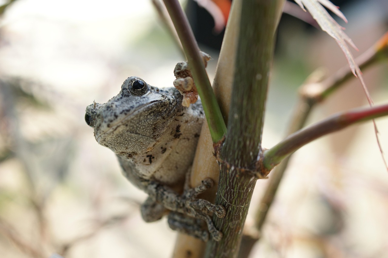
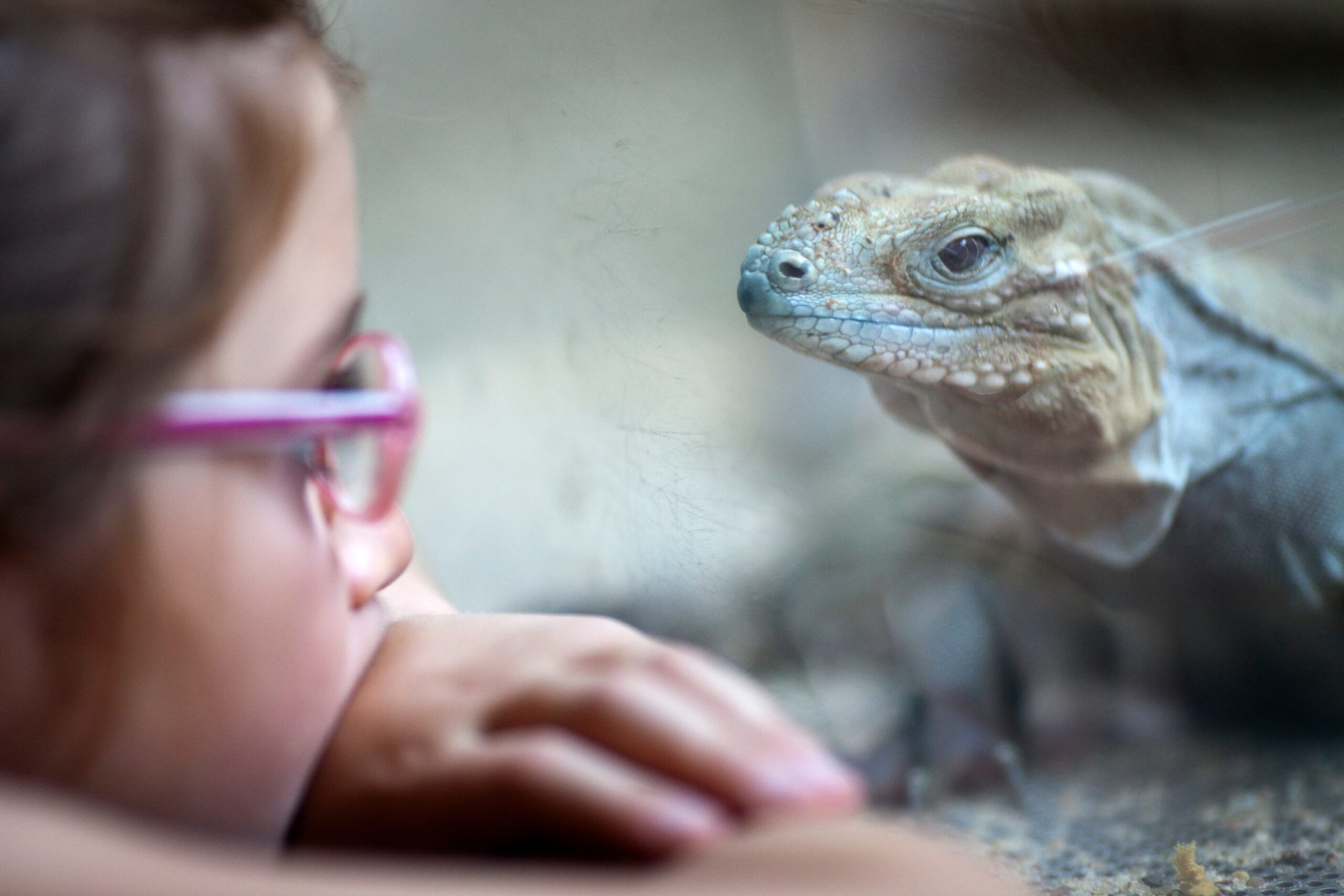
The analysis of estradiol in urine and fecal extracts with the DetectX® Estradiol ELISA Kit offers a noninvasive alternative to serum for hormone monitoring. This is particularly useful in scenarios where blood collection is challenging or not feasible. Urine and fecal estradiol measurements have several applications including human pediatric endocrinology, wildlife and veterinarian science, longitudinal hormone studies, and research that may be sensitive to stress induced by drawing blood.