Assay Principle:
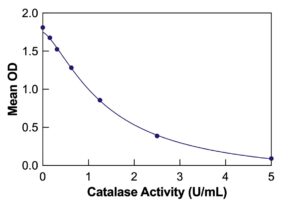
The DetectX® Catalase Colorimetric Activity Kit provides a reliable method for quantitatively measuring catalase activity in serum, plasma, cells, tissues, and RBC/erythrocyte lysates. With a run time of 45 minutes, this colorimetric activity assay enables efficient and accurate analysis. Read the complete kit insert for detailed instructions before starting the assay. The kit includes a Catalase Standard to establish an accurate standard curve.
Protocol Summary:
- Add standards or diluted samples to the provided transparent microtiter plate.
- Introduce Hydrogen Peroxide Reagent to each well, ensuring thorough mixing of reagents.
- Incubate the mixture at room temperature for 30 minutes.
- After this incubation, add the Substrate and the HRP Reagent to each well. Incubate the plate for an additional 15 minutes, allowing the color-generating reaction to occur.
- Use a plate reader to detect the signal at 560nm. Calculate catalase activity using the standard curve.
Background:
Catalase’s main role in the body is to reduce hydrogen peroxide (H2O2). H2O2 is a common reactive oxygen species formed during aerobic metabolism, superoxide formation, dismutation, or as a product of oxidase activity. Excessive hydrogen peroxide and its decomposition product, the hydroxyl radical, are detrimental to cellular components. Rapid removal of hydrogen peroxide is vital for the survival of all aerobically living prokaryotic and eukaryotic cells.
Catalase, an enzyme encoded by a single gene and highly conserved across species, efficiently eliminates peroxide. All tissues in mammals, including humans and mice, express catalase, with high concentrations found in the liver, kidneys, and erythrocytes. Catalase expression is regulated at transcriptional, post-transcriptional, and post-translational levels. Clinicians and researchers typically observe high catalase activity in peroxisomes, various cancers, and conditions like thyroid dysfunction.
The DetectX® Catalase Colorimetric Activity Kit, with its high sensitivity and specificity, is an essential tool for researchers and clinicians studying oxidative stress, cellular defense mechanisms, and diseases related to altered antioxidant enzyme activity.