Retinol Binding Protein (RBP) Multi-Format ELISA Kit
Personal Touch
Here to Help.
Ready to Ship
Most kits in stock.
Easy to Use
Simple protocols.
- Catalog Number K062-H
- Assay Type Competitive ELISA
- Sample Types Serum, Plasma, Urine, Dried Blood Spots
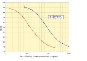
- Sensitivity 5.69 ng/mL (10 µL), 1.36 ng/mL (100 µL)
- Species Human
- Assay Duration 1.5 Hours
- Samples/Plate 38 in Duplicate
- Readout Colorimetric, 450 nm
Assay Principle:
The DetectX® Retinol Binding Protein (RBP) Multi-Format ELISA Kit quantitatively measures RBP in urine, serum, plasma, and dried blood spots. This competitive ELISA has a run time of 1.5 hours. Please read the complete kit insert for more information before performing this assay. Use our provided RBP standard to generate a standard curve for the assay.
Protocol Summary:
- Introduce standards or diluted samples into the included transparent microtiter plate coated with donkey anti-sheep IgG antibody.
- Add RBP peroxidase conjugate and RBP sheep polyclonal antibody to initiate the immunological reaction.
- Incubate the mixture at room temperature with shaking for 1 hour. The reaction inversely correlates with the RBP concentration in the sample.
- Post-incubation, remove excess conjugate and add the TMB substrate, which reacts with the bound conjugate to produce a measurable signal.
- Calculate RBP concentration using a plate reader at 450nm, based on the standard curve.
Background:
Retinol binding protein (RBP) is a critical component in the transport and metabolism of vitamin A (retinol). As a member of a family of proteins that bind small hydrophobic molecules, RBP specifically binds retinol and enables its systemic distribution. This 21 kDa glycoprotein, consisting of 182 amino acids with three disulfide bonds, has a hydrophobic pocket for retinol binding.
RBP is clinically significant as a biomarker for renal function. The protein’s filtration by the glomeruli and subsequent reabsorption in the proximal tubules make it an important indicator of renal health, particularly in heart or kidney transplant recipients, diabetics, and individuals exposed to certain environmental hazards. Moreover, RBP levels correlate with various diseases, including hypertension and certain types of cancer.
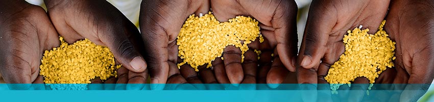
RBP is also notable in monitoring vitamin A deficiency (VAD). The correlation between retinol and RBP in serum indicates that RBP can serve as a surrogate marker for retinol, aiding in the detection of VAD. This is particularly relevant in global health contexts, where nutritional deficiencies and diseases like cystic fibrosis and HIV-1 contribute to the risk of VAD.
The DetectX® RBP Multi-Format ELISA Kit offers a sensitive and specific method for RBP measurement in diverse biological samples. This kit facilitates studies in nutrition, renal health, and disease pathology, contributing significantly to understanding and managing health conditions associated with RBP and vitamin A metabolism.