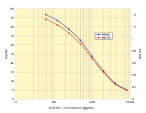
Assay Principle:
The DetectX® DNA Damage ELISA Kit quantitatively measures DNA and RNA oxidized guanosine species in serum, plasma, saliva, urine, fecal extracts, digested DNA, and tissue culture media. This competitive ELISA has a run time of 2.5 hours. Please read the complete kit insert for more information before performing this assay. Use our provided 8-Hydroxy-2’-deoxyguanosine (8-OHdG) standard to generate a standard curve for the assay.
Protocol Summary:
- Introduce standards or diluted samples into the included transparent microtiter plate pre-coated with goat anti-rabbit IgG antibody.
- Add 8-hydroxyguanosine (8-OHG) conjugate and the peroxidase-labeled mouse monoclonal antibody to each well, ensuring thorough mixing.
- Incubate at room temperature, covered and shaking, for 2 hours. The immunological reaction inversely correlates with the 8-OHdG concentration in the sample.
- Post-incubation, remove excess conjugate and add the TMB substrate. The substrate reacts with the bound conjugate, creating a detectable colorimetric signal.
- Utilize a plate reader to measure the signal intensity at 450nm and calculate the 8-OHdG concentration using the standard curve.
Background:
In vivo biological systems frequently generate free radicals and other reactive species, leading to oxidative damage of biomolecules. This damage is counteracted by multiple antioxidant repair mechanisms and the replacement of compromised nucleic acids, proteins, and lipids. Normal metabolism produces intracellular reactive oxygen species (ROS), while environmental factors like UV or ionizing radiation generate extracellular ROS. DNA damage from oxidative stress can disrupt cellular functionality by corrupting genomic information.
Continuous oxidative DNA damage is a significant factor in the age-related emergence of major cancers, including those of the colon, breast, rectum, and prostate. 8-hydroxy-2’-deoxyguanosine (8-OHdG) is a predominant byproduct of DNA oxidation and a biomarker of oxidative stress and carcinogenesis. 8-OHdG is naturally produced during DNA repair processes and is subsequently excreted in urine without further metabolism, making it an effective indicator of oxidative damage at the molecular level.
The DetectX® DNA Damage ELISA Kit is for researchers studying the mechanisms of oxidative stress, DNA repair, and carcinogenesis. It provides a sensitive and specific method to measure 8-OHdG levels in various biological samples, aiding in the investigation of oxidative damage and its implications in health and disease.