Assay Principle:
The DetectX® Glutathione (GSH) Colorimetric Detection Kit offers an efficient method for quantifying GSH levels in a variety of samples. These include whole blood, serum, plasma (EDTA and Heparin), erythrocytes, urine, cell lysates, and tissue. This kit features a rapid run time of only 20 minutes. For best results, read the comprehensive kit insert before starting the assay. A Glutathione standard is included in the kit to facilitate the creation of an accurate standard curve.
Protocol Summary:
- Introduce standards or diluted samples into the clear microtiter plate.
- Add Detection Reagent to each well, ensuring thorough mixing.
- Incubate at room temperature for 20 minutes to enable the color-generating reaction with GSH.
- After incubation, use a plate reader to detect the signal at 405nm and calculate GSH concentration using the standard curve.
Background:
Glutathione (L-γ-glutamyl-L-cysteinyl glycine; GSH), a predominant non-protein thiol in mammalian cells, typically exists in concentrations of 0.5 to 10 mM. As a tripeptide composed of glutamate, cysteine, and glycine, GSH serves as a crucial antioxidant, safeguarding cells from oxidative damage caused by reactive oxygen species.
GSH is enzymatically converted to its oxidized form, glutathione disulfide (GSSG). This process, where GSH acts as an electron donor, is vital in reducing disulfide bonds in cytoplasmic proteins to cysteines. The reduced form of Glutathione is the most common in cells due to the activity of glutathione reductase, which functions continuously and increases in response to oxidative stress.
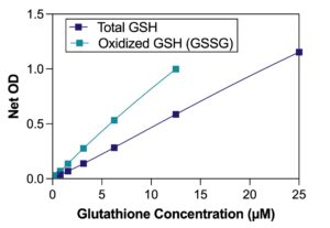
Researchers use the ratio of reduced to oxidized Glutathione within cells as a key indicator of cellular health. This particularly useful in assessing cellular response to oxidative stress and toxicity. Accurate measurement of GSH in its different forms is essential in research areas such as toxicology, pharmacology, and disease studies where oxidative stress is a factor.
The DetectX® Glutathione (GSH) Colorimetric Detection Kit is a valuable biomedical research tool with rapid and sensitive detection capabilities. It enables precise measurement of GSH levels, contributing to a deeper understanding of cellular health and the mechanisms underlying various diseases.