Cortisone
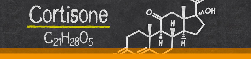
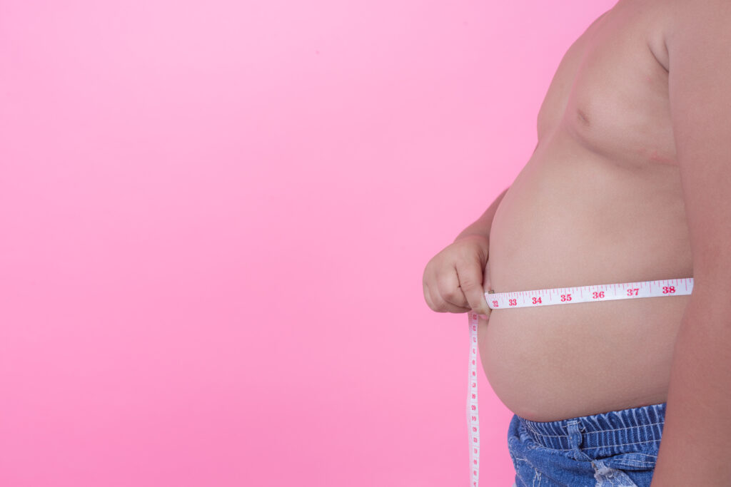
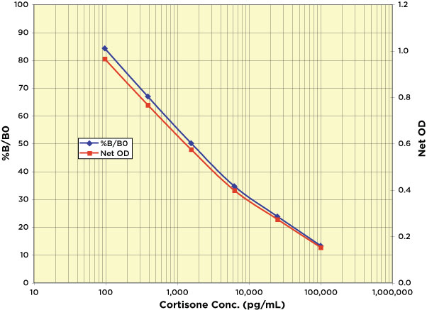
Cortisone was identified by Mason, Myers and Kendall in 1936 as Compound E extracted from bovine suprarenal gland tissue that had the qualitative but not quantitative activity of cortin. The presence of multiple cortin-like compounds led the authors to speculate that the study of Compound E would reveal the nature of cortin. Compound E is now called cortisone and the more active Compound F, cortisol, and the concentrations of these two glucocorticoids vary due to the activity of two 11ß-hydroxysteroid dehydrogenases (11-HSD). While most tissues have the ability to express either enzyme, 11ß-HSD1 is found primarily in the liver where it converts cortisone to cortisol while 11ß-HSD2 is found in tissues such as the kidney where cortisol receptor binding is required. 11ß-HSD2 deactivates cortisol to cortisone, prohibiting receptor activation. This glucocorticoid “shuttle” helps to initiate and regulate the anti-inflammatory response, allowing cortisone to be used as a treatment for a wide range of diseases and conditions. Monitoring the ratio of cortisone:cortisol has applications in diabetes, obesity, metabolic syndrome, osteoporosis, and chronic fatigue syndrome in addition to adrenal diseases. Cortisone and cortisol concentrations exhibit a predictable diurnal pattern and can be measured in extracted dried feces, or in serum, plasma, saliva and urine. Studies have suggested that salivary cortisone may be a good surrogate marker for serum cortisol. Arbor Assays offers both a colorimetric EIA kit and a chemiluminescent CLIA Kit to assess cortisone in a variety of samples.
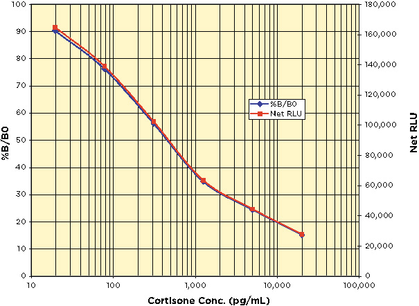
Cortisone Chemiluminescent Immunoassay Kit, K017-C
Related Resources:
Steroid Solid Extraction Protocol
Featured Products
-
 In Stock
Cortisone ELISA Kit
Price range: $436.00 through $1,745.00The DetectX® Cortisone ELISA Kits quantitatively measure cortisone present in a variety of samples.