Validation of Water-Borne steroid Hormones
As human population grows, our species continues to expand into habitats previously occupied by other species around the globe. Unfortunately, we do not make very good neighbors. The impact of human industry and occupation are diverse and devastating. In addition to the obvious physical habitat lost to concrete and paving, there is further harm done by chemical, noise and light pollution, introduction of non-native species, and increased spread of diseases. The emerging field of conservation physiology focuses on how these challenges impact the health of native species. The overall health of an animal population hinges on their ability to successfully reproduce and cope with outside stressors, so measuring stress and reproductive hormones key. Traditionally these endocrine markers have been measured in serum or plasma. However, the act of collecting blood samples from study animals is stressful in and of itself, making it difficult to separate environmental effects from the effects of sample collection. As a result, assays allowing important hormone markers to be measured in non-invasive samples such as urine and feces have become quite important in conservation fields.
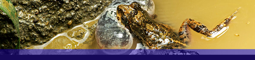
Urine, and feces are great options if you’re working with a terrestrial animal, but can be problematic for aquatic or amphibious species. A recent paper by Baugh et al, offers an alternative by validating the measurement or hormone markers secreted by Túngara frogs into a water bath. Their first step was to determine if the target hormone could be detected in the water bath samples in a meaningful way. So they conducted time course and dose response tests what and without stimulation. In the corticosterone test, male and female frogs received either vehicle, low dose ACTH (to increase corticosterone), high does ACTH. After a 1-hour response time, the frogs were transferred to a series of four 1-hour water baths. After each water bath the water filtered to remove large particulate matter, and stored frozen. Samples were later thawed, extracted (SPE, C18), eluted and dried. Dried samples were reconstituted with 5% ethanol in assay buffer, and assayed using Arbor Assays Corticosterone EIA Kit (K014-H). Progesterone and Estradiol experiments were carried out in similar fashion using only female frogs, stimulation with high or low dose hCG, followed by a 24 hour response time, and a single 60 minute water bath. Samples for progesterone and estradiol were also extracted, eluted and dried prior to assay with Arbor Assays Progesterone EIA Kit (K025-H) and Arbor Assays Estradiol EIA Kit (K030-H). These initial experiments demonstrated that the targeted hormones could be measured in the extracted samples, and that the effects of the stimulation could also be observed with appropriate dose and time responses.
In the next phase, they compared relative values obtained from the water baths to levels measured in plasma samples taken following the final water bath and to whole animal homogenates created after the final water bath. In the corticosterone frogs there no difference was detected between control and ACTH animals in the plasma and homogenate tests. The authors theorized that this animals stress levels may have returned to normal by the time these samples were collected after the end of the water bath. Response to stimulation of estradiol measured in the water baths correlated to responses observed in plasma and in whole animal homogenates. Due to limited plasma samples, progesterone levels were only assessed in the whole animal homogenates. Progesterone levels in the homogenates were very low, and although they did appear to positively correlate with the water bath results, the results were not statistically significant.
Finally, hormone levels in the water bath samples were assessed using HPLC-MS. Somewhat surprisingly, it was discovered at this stage that cortisol was also enriched in the water bath samples. This was unexpected since corticosterone had previously been well established as the primary circulating glucocorticoid metabolite in these frogs. Future studies of water borne steroids in frogs should consider both corticosterone and cortisol to understand what this finding means.
Overall, water-borne corticosterone and estradiol levels increased as expected with the pharmacological stimulators, correlated with levels measured in plasma and homogenates, and were confirmed by HPLC-MS results. Progesterone levels increased moderately in response to the hCG challenge and were detected by HPLC-MS but could not be correlated to levels in the whole animal homogenates. This may be due to complications of either conversion or clearance, and demonstrates that whole body homogenate methods should not be held as the gold standard for understanding endocrine function in small amphibians. The results validate the author’s protocol for collecting, processing and assaying the water bath samples and confirms that this technique can provide a successful, reproducible, biologically relevant, non-invasive means to measure important hormone targets and could potentially be used to provide insight into overall health and viability of individuals in populations of threatened or endangered frogs.